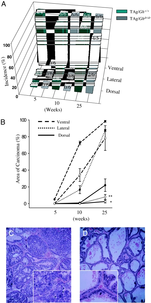
Figure 3.
Prostate carcinogenesis was retarded by disruption of GH signaling in the TAg/Ghdr/dr rat. A, Incidences of microinvasive carcinomas in the two genotypes are shown with the number of rats examined (for example, 2/6 means two of six animals harbored prostatic carcinomas); B, percent area of microinvasive prostatic carcinomas measured using MetaVue image analysis software and expressed as a percentage of total prostate tissue area (•, TAg/Gh+/+; ○, TAg/Ghdr/dr); C and D, representative hematoxylin- and eosin-stained sections of the LP in both genotypes from 25-wk-old rats. The insets in C and D are four times the magnification of the overall image and highlight areas within the larger panels. C illustrates high-grade mPIN and microinvasive carcinomas in TAg/Gh+/+ rats at 25 wk. High-grade mPIN was composed of a markedly increased epithelial cell density with cribriform, micropapillary, or tufting growth patterns, and pronounced cellular atypia. Nuclear chromatin was increased in density, and clumping was common, whereas the basement membrane appeared to be intact. Adenocarcinomas showed microinvasion into the surrounding stroma. Cellular pleomorphism such as nuclear irregularity was much more pronounced in carcinomas than in high-grade mPIN. Prominent nucleoli and apoptotic bodies were often observed. D illustrates normal epithelium and one focus of low-grade mPIN in a TAg/Ghdr/dr rat, which was the predominant phenotype in this animal. *, Significantly different from TAg/Gh+/+ control, P < 0.05; **, P < 0.01.

