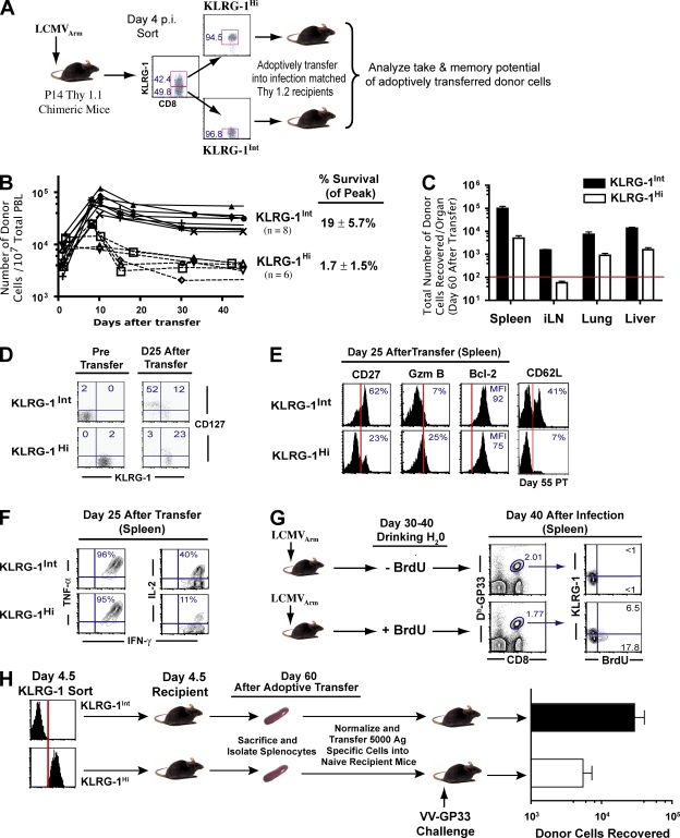
Figure 2.
Heterogeneity in KLRG-1 expression identifies effector CD8 T cells with distinct memory lineage fates. KLRG-1Int effector cells preferentially give rise to long-lived memory cells. (A) KLRG-1Int and KLRG-1Hi cells were sorted from B6 mice containing 106 Thy1.1+ P14 cells 4 d after LCMV infection. Equal numbers (∼106) of sorted cells were transferred into infection-matched Thy1.2 recipients. The numbers indicate percentages of corresponding gated populations. (B) Expansion and contraction of adoptively transferred KLRG-1Int and KLRG-1Hi cells was longitudinally assessed in the blood of recipient mice by staining for the Thy1.1+ marker. (C) Long-term survival of the sorted donor KLRG-1Int and KLRG-1Hi effector cells was determined by enumerating the total number of donor cells in the spleen, lymph node, lung, liver, and blood ∼60 d after adoptive transfer. The horizontal red line indicates the lower limit of detection for absolute numbers of antigen-specific cells. Mean data from six to eight mice are presented, and error bars represent SEM. (D and E) Detailed phenotypic characterization of surviving KLRG-1Int and KLRG-1Hi donor cells in spleens at memory. Percentages of donor cells expressing the indicated markers are depicted. The MFI of Bcl-2 expression is presented. Vertical red lines indicate the negative expression gate for the respective markers. (F) Cytokine production by donor cells at day 25 after transfer. Production of intracellular cytokines (IFN-γ, TNF-α, and IL-2) in Thy1.1+ donor cells was evaluated after 5 h of stimulation with GP33-41 peptide in the presence of BFA in vitro. Percentages of donor cells producing IFN-γ, TNF-α, and IL-2 are depicted in the respective histograms. (G) Homeostatic proliferation of KLRG-1Int and KLRG-1Hi memory cells was assessed in LCMV-infected B6 mice. Immune mice were fed BrdU in their drinking water between days 30 and 40 p.i. Subsequently, BrdU incorporation in KLRG-1Int and KLRG-1Hi splenic DbGP33–specific memory cells was evaluated by flow cytometry. The numbers indicate percentages of corresponding gated populations. (H) Recall proliferation and boosting ability of KLRG-1Int and KLRG-1Hi memory cells. KLRG-1Int and KLRG-1Hi P14 cells were adoptively transferred into infection-matched mice; 30 d later, equal numbers of surviving memory cells (Thy 1.1+) from each group were transferred into Thy1.2+ naive mice, which were infected i.v. with 2 × 10⁶ PFU VV-GP33. Donor cells recovered from the spleen at day 60 after challenge are plotted. Mean data from six mice are presented, and error bars represent SEM.

