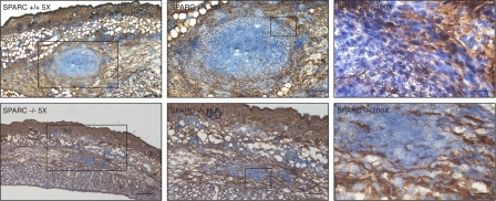
Figure 2.
Collagen IV staining reveals disorganized collagen deposition and more scattered infiltrate in SPARC−/− mice. SPARC+/+ (top row) and SPARC−/− (bottom row) mice were injected in the skin with 107 CFU of attenuated S. typhimurium SL3261 AT. Immunohistochemical staining with anti–collagen IV (brown) and counterstaining of nuclei with hematoxylin (blue) is shown on sections from mice killed 5 d after infection. Numbers show the original magnification of the sections. Boxes represent the magnified areas in the adjacent right images. Bars: (left) 50 μm; (middle) 25 μm; (right) 5 μm.

