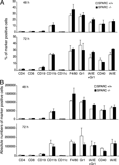
Figure 3.
Inflammatory cell infiltrate composition is very similar in SPARC+/+ and SPARC−/− mice. SPARC+/+ and SPARC−/− mice were injected intradermally in the back skin with 107 CFU of attenuated S. typhimurium SL3261 AT 48 and 72 h later, the sites characterized by an inflammatory reaction were excised, and cells were isolated by collagenase treatment. Cells were stained with fluorescent antibodies and analyzed by flow cytometry. (A) Y axes show percentage of marker-positive cells, x axes show the analyzed markers. (B) Y axes show total numbers of marker positive cells, x axes show the analyzed markers. Error bars represent the SD of three independent mice. One of three independent experiments is shown. SPARC −/−, black bars; SPARC+/+, white bars.

