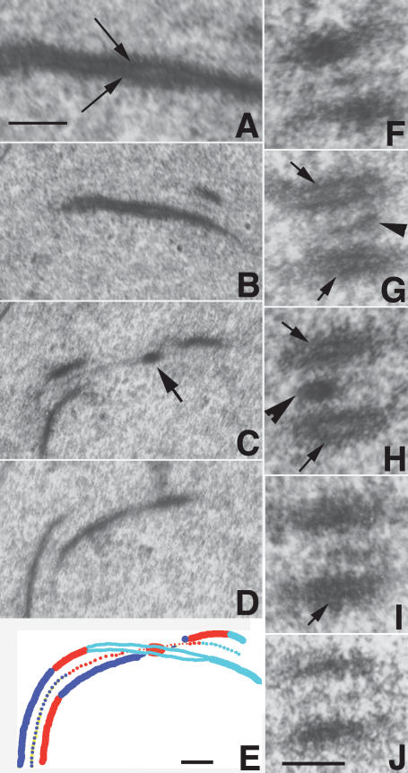
Figure 6.
Electron micrographs of wild-type SCs. (A) LE in longitudinal section. Arrows point to the local separation of the two chromatids. (B–D) Three consecutive serial sections of a SC segment. The recombination nodule (arrow in C) is located at the dual LE site (B) enlarged in A. (C,D) Note that LEs adjoining the nodule are single units. (E) Reconstruction drawing of B–D (B in cyan, C in red, D in blue) showing that the SC twists at the nodule site. (F–J) Five consecutive oblique/cross-sections through a SC segment exhibiting local separation of sister chromatids (arrows) on both homologs (G–I) at the nodule site (arrowhead in G,H); in contrast, in the two adjacent sections (F,J), LEs are single units. Bars, 100 nm.

