Figure 5.
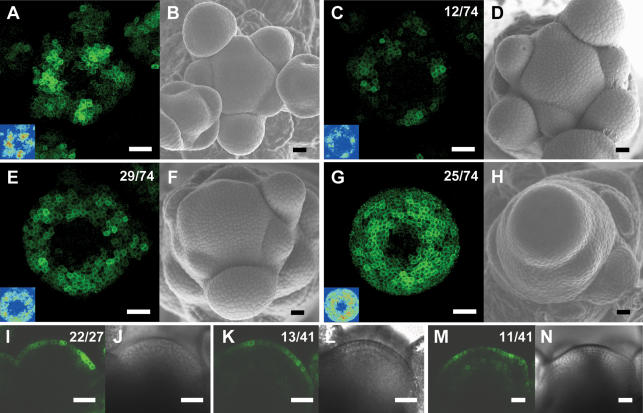
The pattern of DR5∷GFP expression is altered in aux1 lax1 lax2 triple mutant inflorescence meristems. (A) Maximal projection of transversal confocal scans of a wild-type meristem expressing DR5∷GFP. (Inset) LUT signal intensity monitor. (Blue) Low intensity; (red) high intensity. (B) Scanning electron microscope image of the same wild-type meristem as in A. (C,E,G) Maximal projections of transversal confocal scans of DR5∷GFP-expressing aux1 lax1 lax2 triple mutant meristems displaying increasingly severe phenotypes. (Inset) LUT signal intensity monitor. Representative meristems showing smaller and weaker peaks (C), broader and less defined peaks (E), or expression throughout the peripheral zone (G). (D,F,H) Scanning electron microscope images of the same meristems as in C, E, and G, respectively. (I) Longitudinal confocal section through a DR5∷GFP-expressing wild-type meristem showing no GFP expression in inner layers, except below initiation sites. (J) Transmission light picture of the wild-type meristem shown in I. (K,M) Longitudinal confocal section through two aux1 lax1 lax2 triple mutant meristems, showing very weak (K) or weak (M) DR5∷GFP expression in inner layers. (L,N) Transmission light pictures of the meristems shown in K and M. Bars, 25 μm. Numbers indicate the number of occurrences over the total number of observations.

