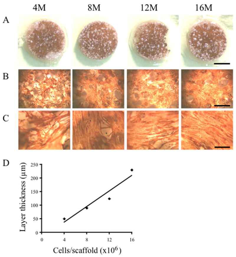
Fig 1. Differentiation of human skeletal myoblasts to myofibers on PLG scaffolds.

Various numbers (4, 8, 12 or 16 million cells, indicated on top of figure) of human myoblasts were seeded in 0.1 mg/ml collagen ECM and differentiated to myofibers on 13 mm PLG scaffolds containing 10% microspheres and prefilled with 0.1 mg/ml collagen ECM. After 5 days in differentiation medium, cells were stained for the expression of sarcomeric tropomyosin (brown color). Myofibers were short and not aligned in any specific direction. (A) Picture of entire scaffold. Scale bar represents 5 mm. (B) 2.5× Objective, scale bar represents 1 mm. (C) 10× Objective, scale bar represents 0.25 mm. (D) Cell layer thickness was measured on thin sections of paraffin embedded scaffolds and is linearly dependent on the initial number of seeded cells.
