
Fig 4. Cell survival as a function of ECM and scaffold composition.
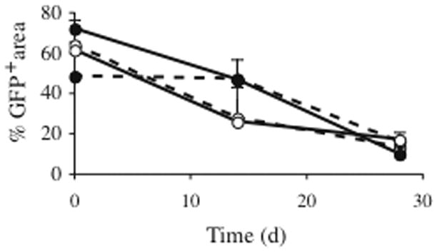
Scaffolds (4.2 mm diameter) composed of either 100 % microspheres (solid lines) or 50 % microspheres (dotted lines) and seeded with 1.5 million human GFP human myoblasts were implanted subcutaneously in the back of NOD/SCID mice. Cells were embedded in a fibrin ECM (open circles) or collagen ECM (filled circles). Datapoints represent GFP area as quantified by confocal analysis pre-implant and after 2 or 4 weeks in vivo. At 2 weeks in vivo, myofibers embedded in a collagen ECM on average covered more area than myofibers in the fibrin ECM; however this difference was not statistically significant. At 4 weeks, GFP area for all conditions was 10–17% (22±2% survival when area was normalized to preimplant GFP area), with no statistical differences for the tested conditions.