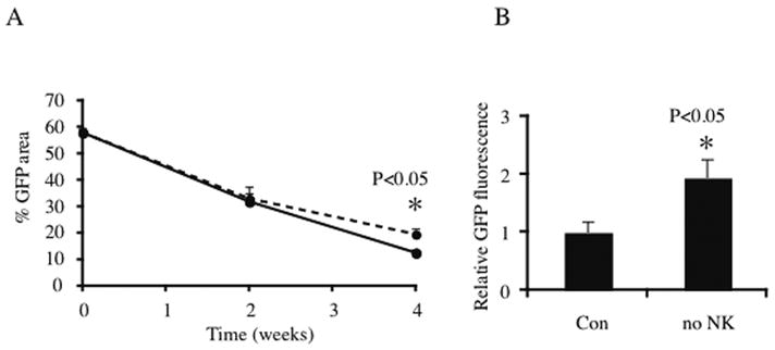
Fig 6. Effect of NK cell depletion on cell viability.

100 % microsphere, 7mm diameter PLG scaffolds were seeded with 4 million GFP myoblasts in a fibrin ECM and implanted subcutaneously in NOD/SCID animals. (A) GFP area on the scaffolds was quantified by confocal microscopy pre-implant and after 2 or 4 weeks in vivo. In the NK cell depleted group (dotted line), animals received intravenous injections of anti-ASGM1 antibody, while control group (solid line) received an isotype serum control. No significant difference was apparent at week 2, but at week 4, myofiber survival in the NK depleted group was significantly (p<0.05) higher than control. (B) Scaffolds in the NK depleted animals contained 2-fold as much GFP protein compared to the control group (p<0.05) after 4 weeks in vivo.
