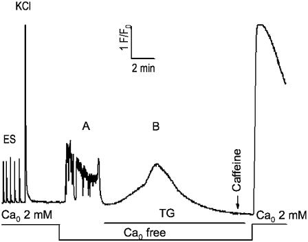
FIGURE 8.
Complete SR Ca depletion protocol for α2δ1 knockdown myotubes. Cells were loaded with Fluo-4 as described in Materials and Methods and tested for responses to electrical pulses and K+ (40 mM) application in Ca-replete medium ( 2 mM). The extracellular solution was exchanged with one lacking added Ca2+ (
 free); it elicited spontaneous activity in most cells. TG (200 nM) was then perfused onto the cells in the
  -free medium until the new baseline was established (typically <10 min) and depletion of SR Ca tested with a 20 mM caffeine challenge. The presence of SOCE was then tested by perfusion of 2 mM Ca2+ medium. This protocol completely depleted SR Ca in >95% of all myotubes exhibiting EC coupling. Wt myotubes were indistinguishable from the α2δ1 knockdown myotubes shown here.

