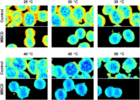
FIGURE 5.
Effects of MBCD treatment on laurdan GP at multiple temperatures. Two-photon images of erythrocytes were gathered at 25°C, 30°C, 35°C, 40°C, 45°C, and 50°C as explained in Materials and Methods. Control cells are shown on the top half of each panel with MBCD-treated cells below. Laurdan GP values range from −0.50 (blue) to 0.75 (red).

