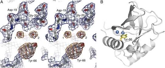
FIGURE 1.
(A) Stereo view of electron density maps of the Tyr-66 protein at room temperature after molecular replacement, contoured over the final refined coordinates. The Tyr side chain and water molecules 1 and 2 are unambiguously shown in the 1.25 σ 2Fo–Fc (blue) and 3.0 σ Fo–Fc (orange) electron density. (B) Ribbon representation of the final structure of the variant with Tyr-66 at room temperature with water molecules 1 and 2 displayed.

