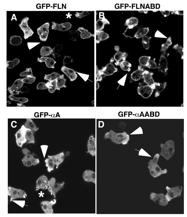
Figure 3.
The localization of filamin and α-actinin derived probes in non-polarized cells. (A and B) When expressed in vegetative cells the localization pattern of GFP-FLNABD (B) was similar to that of GFP-FLN (A). Both probes localized strongly to the peripheral cortex, the leading edge of motile cells (arrowheads), new pseudopods (arrows) and macropinocytic cups (*). (C and D) When expressed in vegetative cells both GFPα-A (C) and GFP-αAABD (D) are present in new protrusions (arrows) and at the leading edge of motile cells (arrowheads) and to macropinocytotic cups*, but not in the peripheral cortex.

