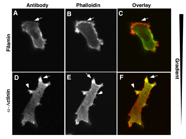
Figure 8.
Immunostaining of polarized cells. AX2 wild type cells were fixed and stained with affinity purified antibodies to either filamin or a-actinin (green). The cells were then counterstained with rhodamine phalloidin (red) to visualize total F-actin. In some cells, filamin was missing from the front of the cell, in a region that was clearly stained with phalloidin (A-C). α-actinin was localized to the leading edge protrusions but relatively absent from the rear of the cell. Both localization patters mirror the results found with the GFP probes.

