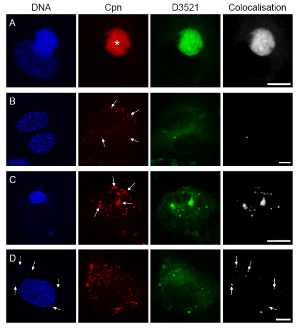
Figure 6.
Detection of BODIPY FL C5-Ceramide. HAEC were infected with 10 IFU/cell for 24 h or 48 h and subsequently incubated with D3521 for 24 h. Controls were additionally treated with chloramphenicol. Cells were stained with anti C. pneumoniae-MOMP (red), D3521 (green), DAPI (blue) and colocalization of D3521 and MOMP was calculated (white). A: Infected HAEC 72 hpi contain a MOMP positive inclusion (asterisk). B: Spot-like infected healthy HAEC 48 hpi display a number of MOMP positive spots (arrows) of which only very few colocalize with the ceramide signal. Hence, 24 h after ceramide supply D3521 colocalizes with the MOMP signal in the inclusion but not in the spots of healthy infected host cells (A, B). C: Aponecrotic spot-like infected cell 48 hpi display a high number of MOMP positive spots resp. aggregates (arrows). The majority of the C. pneumoniae-spots colocalize with the D3521 signal, reflecting metabolic activity. D: Chloramphenicol treated infected HAEC 48 hpi display a normal nuclear morphology and carry a variety of C. pneumoniae-spots. Few of them colocalize with D3521. Note that the D3521 signal occurs exclusively in spots displaying a DAPI labelling (arrows). Scale bar; 10 μm.

