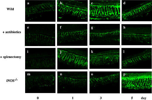
Fig. 6.

Histological examination of the expression of IgA in the colon. At the indicated times after BDL, colon specimens were frozen, cut into thin sections, treated with anti-IgA antibody and then stained with FITC-conjugated second antibody. Other conditions were the same as in Fig. 1. Scale bar = 50 µm.
