
Fig. 8.
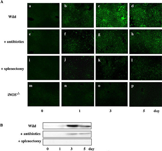
Effect of BDL on hepatic expression of iNOS. At the indicated times after BDL, liver specimens were incubated with anti-iNOS antibody and then stained with FITC-conjugated second antibody (A). Expression of iNOS was determined by Western blot analysis (B). Equal amounts of protein (30 µg/lane) were subjected to a 7.5% SDS-PAGE, and the electrophoresed proteins were transferred onto Immobilon-P membranes, and analyzed using specific anti-iNOS antibody. The bands with 130 kDa responsible for iNOS were seen in liver specimens from BDL mice. Data show one typical result out of 3 representative experiments. Scale bar = 100 µm.