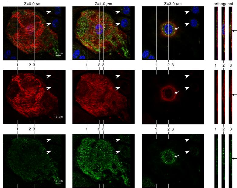
Figure 2.
Krp1 (green, lower row) and caveolin-3 (red, middle row) localization in a single cultured mouse cardiomyocyte by confocal microscopy. DAPI labeling of the nucleus is also shown in the blue channel of the composite images (top row). Confocal imaging at planes 0.0, 1.0, and 3.0 μm above the adherent ventral surface of the cardiomyocyte are shown, as indicated. Orthogonal views through the cell are shown at the right, and their positions in the x-y plane are indicated in the adjacent micrographs. The caveolin-3 staining marks the ventral and dorsal surfaces of the cell. Punctate patches of Krp1 are clearly localized within the cytoplasm (arrows), in between the cell boundaries marked by caveolin-3.

