Figure 5.
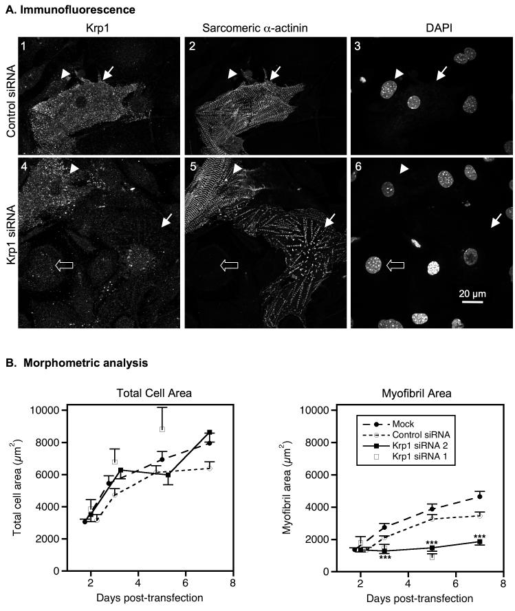
Krp1 knockdown decreases mature myofibril content. (A) Cells were fixed and stained for Krp1 and sarcomeric α-actinin 5 days after transfection with control siRNA (top row) or Krp1 siRNA (bottom row). Nuclei were counterstained with DAPI. Images are representative of cells evaluated from three independent cultures. Control cardiomyocytes stain positively for Krp1 and contain sarcomeric α-actinin organized into mature striations (panels 1-3, arrow). Decreased levels of Krp1 after siRNA treatment are associated with low levels of mature myofibrils (panels 4-6, arrow), while some cardiomyocytes retain normal Krp1 levels and exhibit normal striations (panels 4-6, arrowhead). Neighboring fibroblasts exhibit only background staining for Krp1 (panels 1-3, arrowhead; panels 4-6, open arrow). (B) Morphometric analysis of total cardiomyocyte areas (left) and mature myofibril areas (right) versus time. Each point represents the mean and SEM of 25-58 cardiomyocytes from at least 3 independent experiments, with the exception of the Krp1 siRNA 1 data where each point is the mean value of 10-23 cardiomyocytes from 1-3 independent experiments. Krp1 siRNA halts myofibril accumulation after 2 days, but cardiomyocyte spreading is unaffected. p-values compared to mock-transfected controls: ***p <0.001.

