Figure 8.
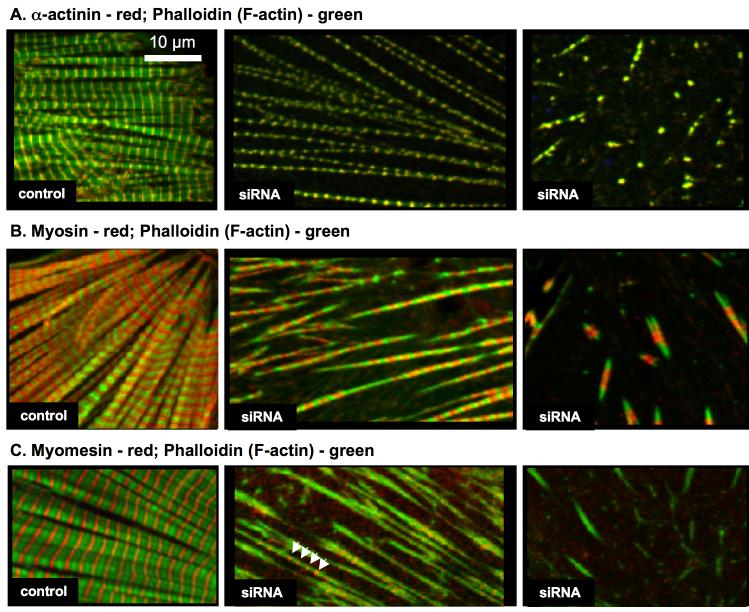
Organization of sarcomeric components after Krp1 knockdown. Cardiomyocytes were fixed five days post-transfection and double stained with phalloidin to visualize actin filaments (green) and antibodies for either sarcomeric α-actinin (A), muscle myosin (B), or myomesin (C) (red). Examples of control cardiomyocytes (left panels) and cells transfected with Krp1 siRNA 1 (center and right panels) are shown. In control cardiomyocytes, the sarcomeric proteins are organized into mature, well-aligned myofibrils. In contrast, after Krp1 knockdown cells contain sparse, separated fibrils or very short fibrils, which still contain α-actinin, actin and myosin organized in the same banding pattern observed in mature myofibrils. Myomesin is present in the longer fibrils (arrowheads), but is often absent from the shorter fibrils (C, center and right panels, respectively).

