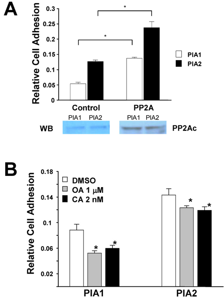
Figure 4. PP2A regulates αIIbβ3-dependent adhesion under motion in vitro.

(A) PlA1 and PlA2 CHO cells were transiently transfected with PP2Ac (PP2A) or control vector (Control) and assayed for adhesion on fibrinogen-coated plates 36 hours post-transfection. The results are representative of three independent experiments. The presented data are the mean ± SD of triplicates in one experiment where (*) represents P < 0.01 (n = 3). Western blot showed the cell expression level of PP2A. (B) PlA1 and PlA2 CHO cells were treated with either okadaic acid (OA), or calyculin A (CA), or with DMSO control (DMSO) for 30 min, and then analyzed by adhesion assay on fibrinogen-coated plates. The results are representative of three independent experiments. The presented data are means ± SD of triplicates in one experiment. The difference between inhibitor treated vs DMSO-treated cells is significant where (*) represents P < 0.01, n = 3.
