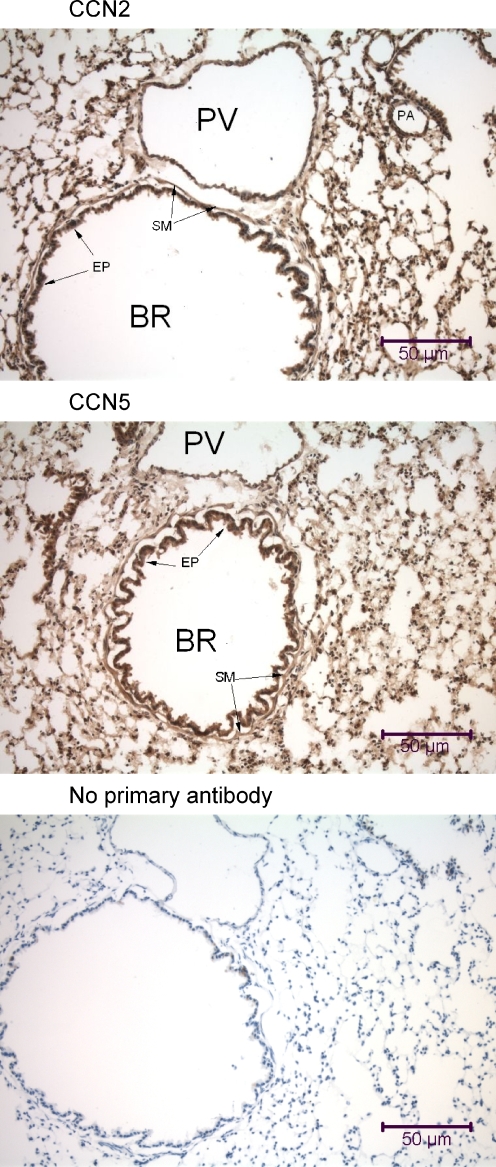
Fig. 2.
Immunohistochemical analysis of the mouse lung using anti-CCN2 and anti-CCN5. Lung samples were obtained from a mouse that was fixed by intracardial perfusion immediately post-mortem. In each panel is a transversely-oriented 7 μm lung tissue section that reveals a large bronchiole (BR) with a prominent lumenal columnar epithelium (EP) surrounded by a thin layer of circumferential smooth muscle (SM), large thin-walled pulmonary veins (PV), some small pulmonary arteries (PA), and numerous alveolar air sacs. All types of cells in the mouse lung demonstrate some nuclear localization of both CCN2 and CCN5

