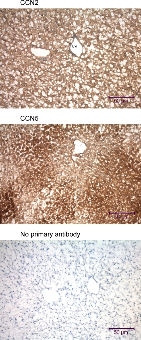
Fig. 6.
Immunohistochemical analysis of the mouse liver using anti-CCN2 and anti-CCN5. Liver samples were obtained from a mouse that was fixed by intracardial perfusion immediately post-mortem. The tissue sections (7 μm transverse) display cords of hepatocytes surrounded by sinusoids, large central veins (CV), and smaller portal vein branches

