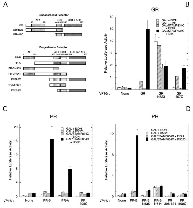
Fig. 6.

Regions of PR and GR interacting with STAMP in two-hybrid assays. (A) Cartoons show regions of GR and PR that are retained in each of the constructs used below, with the numbers above each structure indicate the amino acid position of each domain boundary. Plasmid names are that of the receptor followed by the sequence retained, with the letters N and C designating the N- and C-terminal residues of the receptor respectively. (B-D) Cos-7 cells were transiently transfected with GAL (57 ng) or GAL/STAMP834C (80 ng) plus VP16 fusions of GR segments (molar equivalent to 86 ng VP16/GR; B) of PR segments (molar equivalent to 1 ng VP16/PR-B; C&D) and 100 ng of FRLuc reporter ± 1 μM Dex (B) or 20 nM R5020 (C&D). The average relative luciferase activities from three independent experiments were determined and plotted as in Fig. 5.
