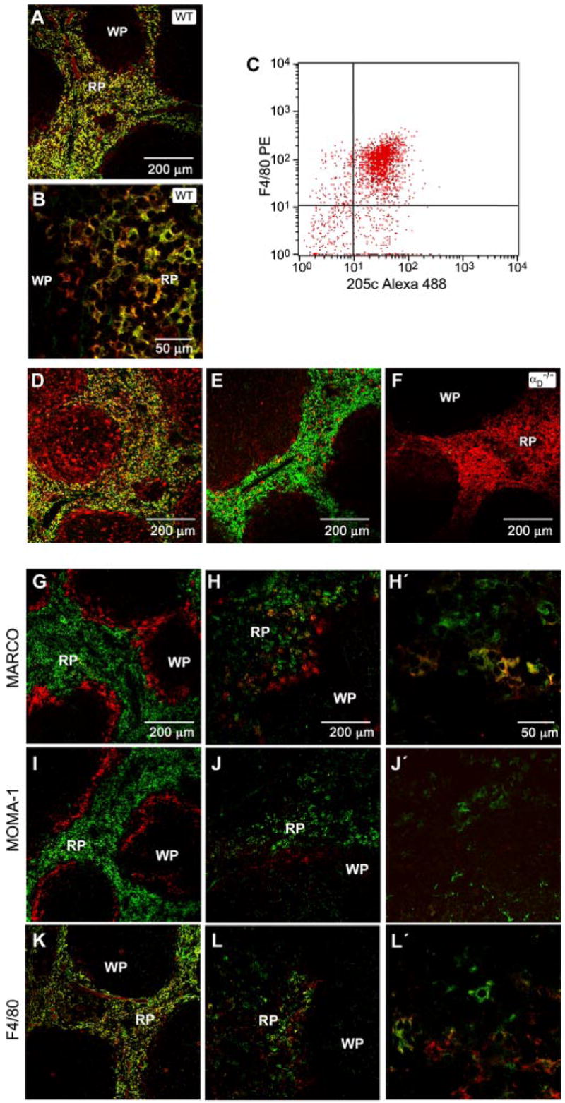
FIGURE 3.

αDβ2 is expressed by a unique population of splenic red pulp macrophages that is altered in P. berghei infection. Frozen sections of freshly isolated spleen from WT mice were costained using anti-αD mAb (mAb 205c, green fluorescent labeling) and mAb against F4/80 (A and B), macrosialin (D), or αm (E; red labeling). F, Spleen sections from αD−/− mice were costained for αD and F4/80 as in A and B. The staining patterns shown are representative of tissue from multiple WT and αD−/− mice. C, Splenocytes were selected based on binding of PE-labeled anti-F4/80 and examined for expression of αD by flow cytometry using mAb 205c conjugated to Alexa 488. A similar pattern was seen in two additional experiments. G–L, Frozen sections of spleen from control (G, I, and K) or P. berghei-infected (7 days postinfection; H, J, and L) WT mice were costained for αD and MARCO, MOMA-1, or F4/80. In each set of panels, αD was detected by green fluorescence and the second marker was detected by red fluorescence. The right panels (H′, J′, and L′) show higher magnification detail. The staining patterns were similar in tissue from three infected animals. RP, Red pulp; WP, white pulp.
