
Fig. 3.
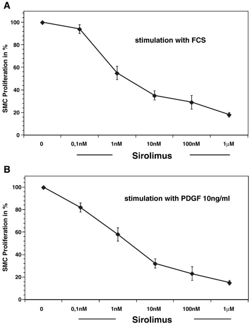
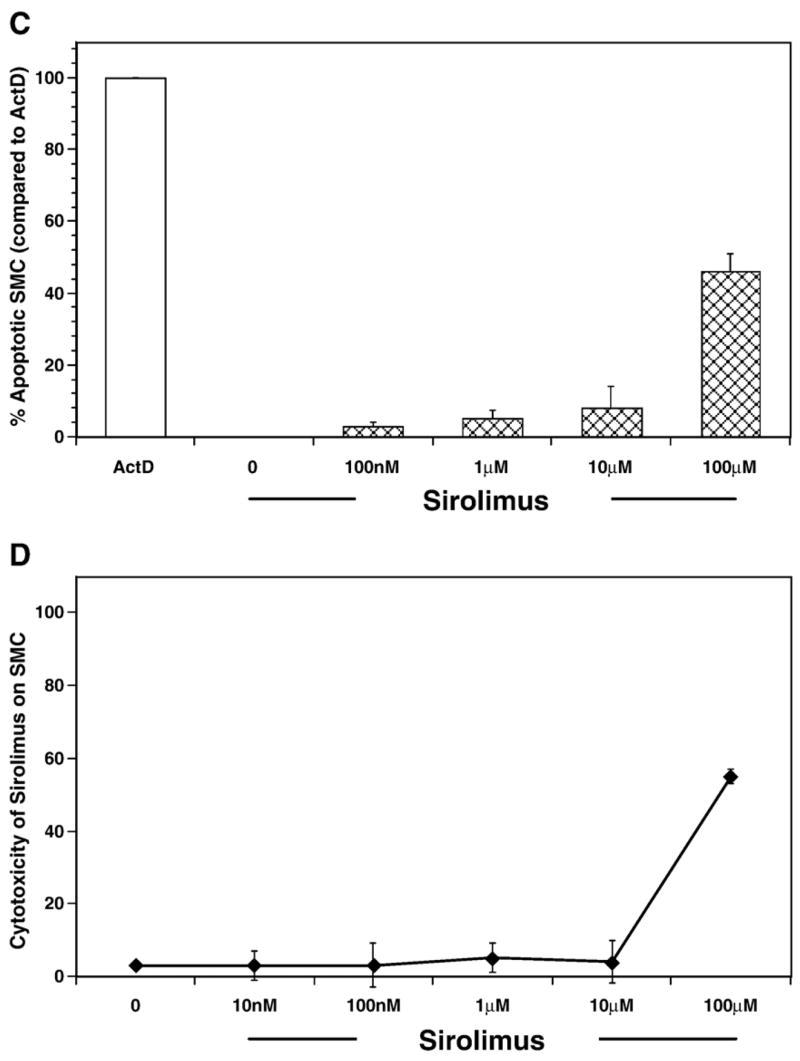
Sirolimus blocks smooth muscle proliferation independent of cell death. (A, B) Bar graph showing the concentration response for sirolimus-induced inhibition of cultured rabbit vascular smooth muscle cell (VSMC) proliferation. Cells were treated for 10 min with either vehicle or increasing concentrations of sirolimus and subsequently stimulated with either 10% FCS (A) or PDGF-α (10 ng/ml) (B). The results are the mean±SEM for 8 experiments. (C) Bar graph showing the percentage of cells that are apoptotic in actinomycin D or sirolimus treated smooth muscle cells. (D) Bar graph showing the cytotoxic effect of sirolimus on cultured rabbit aortic smooth muscle cells. The results are the mean±SEM of six independent experiments.