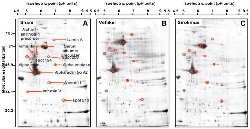
Fig. 4.
Sirolimus differentially blocks the expression of structural proteins within the injured vessel wall. Vessels were left intact (sham) or exposed to angioplasty where an intramural injection of sirolimus (PTA+sirolimus) or vehicle (PTA+vehicle) was delivered through the balloon catheter during the angioplasty procedure. The vessels were collected after 3 weeks, proteins were harvested and normalized, and the proteins were separated by 2-D gel electrophoresis. Proteins on the gel were identified by silver stain as described in Materials and methods. (A) Sham (B) PTA+vehicle. (C) PTA+sirolimus. Size of original gel: 16×16×0.1 cm. Circled spots in panel B are proteins that were increased in angioplastied vessels compared to uninjured vessels (A). For comparison, the same areas are circled in panel C. Proteins that were eventually identified by nanospray tandem mass spectrometry (see Fig. 5 below) are labeled accordingly. This figure is representative of 5 independent experiments for each group.

