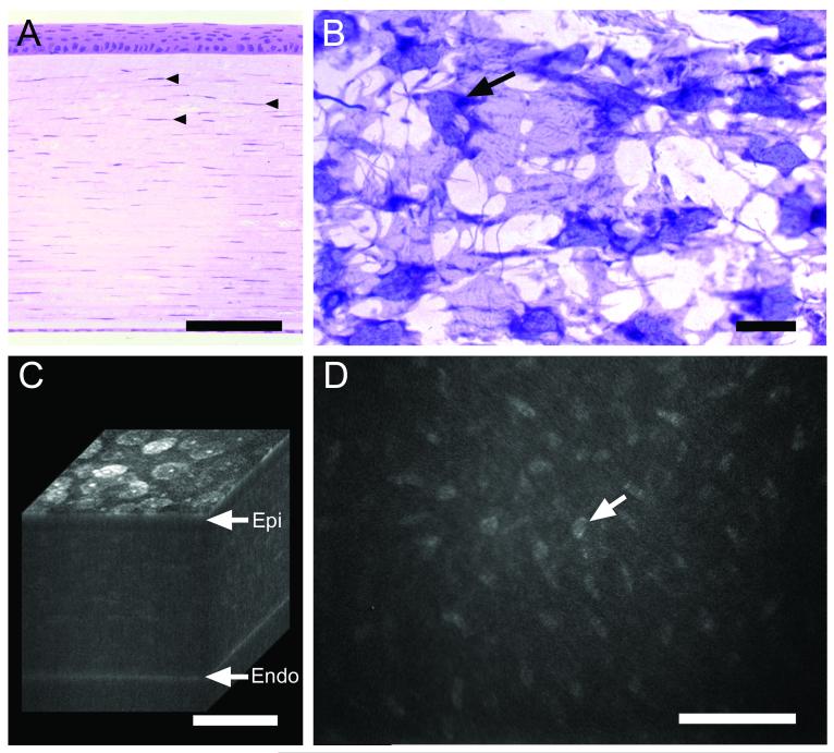
Fig. 1.
Normal rabbit cornea as shown by histology (A and B) and in vivo confocal microscopy (C and D). (A) Cross-section stained with hematoxylin & eosin showing anterior stratified corneal epithelium overlying corneal stroma with keratocyte nuclei (arrowheads) and posterior corneal endothelium. (B) Coronal section through the corneal stroma stained with gold-chloride showing keratocyte cell bodies and nuclei (arrow). (C) 3-D reconstruction of confocal images through a living rabbit cornea showing surface epithelial cells (Epi), underlying stroma and posterior corneal endothelium (Endo). (D) 2-D in vivo confocal image taken from the 3-D data set through the corneal stroma showing light scattering from the keratocyte nuclei. Bar = 100 μm.

