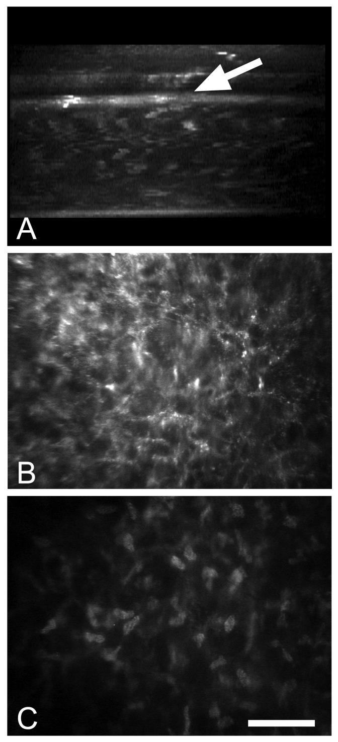
Fig. 4.
In vivo confocal microscopy of a patient following intrastromal implantation of a corneal lens. (A) X-Z projection through the 3-dimensional data set showing the anterior placement of the intracorneal lens (arrow) and the associated increased light scattering at the posterior margin of the lens. (B) X-Y plane taken from the 3-dimensional data set just posterior to the intracorneal lens showing marked light scattering from corneal keratocytes. (C) X-Y plane taken from the 3-dimensional data set further posterior within the normal stroma showing light scattering limited to keratocyte nuclei. Bar = 100 μm.

