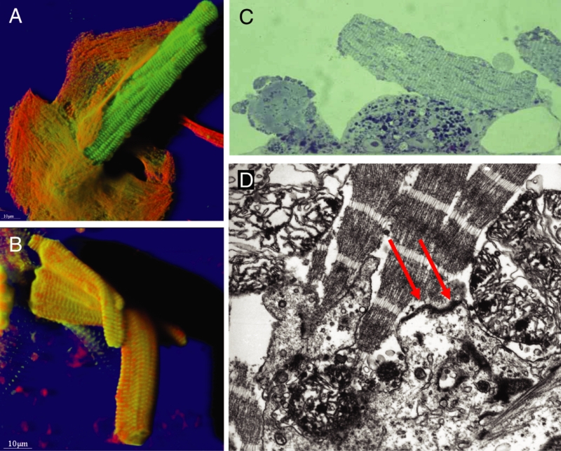
Figure 5).
Different modes of attachment of ‘second-floor’ cells to underlying spreading myocytes. Red represents actin and green represents alpha-actinin. A Shadow projection. The second-floor cell is attached with only a small tip of its cytoplasm to the underlying cell (top left corner). B Shadow projection. Attachment by engulfment of one second-floor cell by spreading myocytes. C Light microscopy of semithin sections shows the typical localization of a second-floor cell on a healthy, dedifferentiating cardiomyocyte (original magnification ×400). D Electron micrograph showing still existing attachment points between two myocytes (arrows), one living (bottom) and one dead (top) (original magnification ×39,000)

