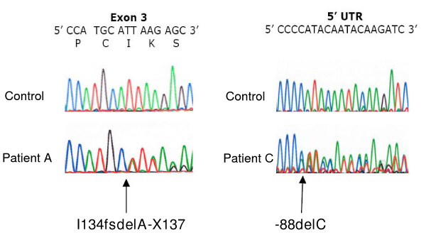
Figure 1.
Electropherogram scans showing the novel heterozygous CAV1 frameshift mutations in the lipodystrophy patients. The left half of the figure shows a portion of CAV1 exon 2 from genomic DNA of a control subject and Patient A. The right half of the figure shows a portion of CAV1 5' untranslated region (5'UTR) from genomic DNA of a control subject and Patient C. For each tracing, normal nucleotide sequence is shown in the top line of letters, with single letter amino acid codes and codon numbers beneath for exon sequence. The position of each inserted nucleotide is indicated by the arrows for the respective mutations I134fsdelA-X137 and -88delC.

