
Figure 7.
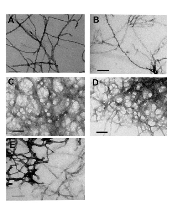
The morphology of amyloid fibers formed from protein containing pathological versus non-pathological repeat lengths is distinct. Unseeded fiber formation reactions (5 μM) were imaged by TEM after 16 hours. The length of scale bar represents 100 nm. (A) Sup35NM and (B) SP5NM have uniform dispersed fibers. (C) SP8NM, (D) SP11NM and (E) SP14NM have clumped fibers.