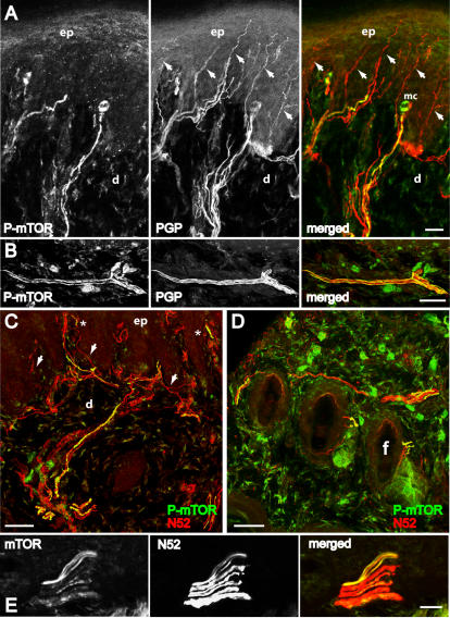
Figure 1. Distribution of mTOR immunoreactivity in peripheral sensory nerve fibers in the skin.
Confocal images of 40 µM thick frozen sections cut perpendicular to the skin surface of the rat hindpaw. A, B, Colocalization of phospho-mTOR (green) and nerve fibers marker PGP (red), a general marker of nerve fibers, in footpad of glabrous skin. Arrows indicate PGP- positive fibers not double-labelled. C, D, Colocalization of phospho-mTOR (green) and myelinated fiber marker N52 (red) in footpad of glabrous skin (C) and hairy skin (D). Arrows in C indicate N52- positive fibers not double-labelled. Asterisks (*) indicate double-labelled fibers in the area of Meissner corpuscles (sensory receptors within dermal papillae). E, Colocalization of phospho-mTOR and N52 in the dermis of the glabrous skin. In A, B and E the single staining for each antibody and the merged image are shown from left to right. In A-E double staining appears in yellow. mc, Meissner corpuscles; ep, epidermis; d, dermis; f, follicle. A, E, single focal planes; B-D, merge of 22–24 z-focal planes (20–21 µm depth). Scale bars, A-E: 50 µm.

