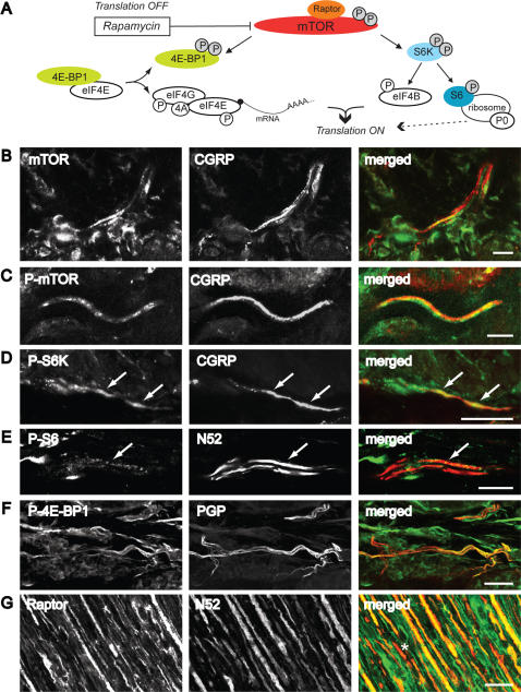
Figure 2. Translation-related factors are localized in peripheral nerve fibers and terminals in the skin.
A, Diagram of mTOR signalling pathways and translational modulation. P, phosphorylation [24], [33], [34], [80]. B-F, Confocal images of 40 µM thick sections cut perpendicular to the plantar skin surface of the rat hindpaw. B-D, Colocalization of mTOR (B) phospho-mTOR (C) or phospho-S6K (D) and CGRP positive fibers in the dermal glabrous skin. E, Colocalization of phospho-S6 and N52 in the dermis of glabrous skin. F, Colocalization of phospho-4E-BP1/2 and PGP in the dermal glabrous skin. G, Colocalization of raptor and N52 in the sciatic nerve. In B-G, the single staining for each antibody and the merged image are shown from left to right. Double staining appears in yellow. White arrows indicate double-labelled fibers. Asterisk (*) indicates N52- positive fibers not double labelled. B, E single focal planes; C, 8 z-focal planes (2.3 µm); D, merge of 14 z-focal planes (8.5 µm depth); F, merge of 59 z-focal planes (17 µm depth); G, 28 z-focal planes (11 µm). Scale bars, B-E, G: 25 µm; F: 10 µm.

