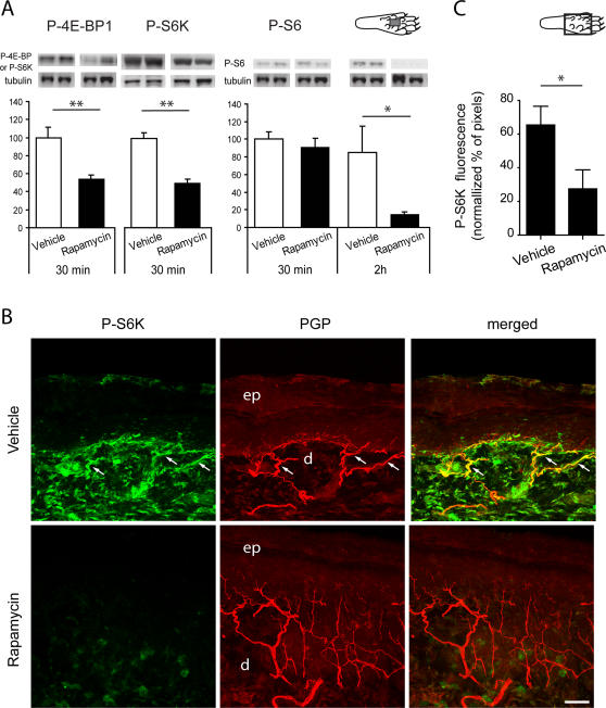
Figure 3. Rapamycin decreases phosphorylation of downstream targets of mTOR in the skin.
A, Immunoblots probed with anti-phospho-4E-BP1/2, anti-phospho-S6K and anti-phospho-S6 antibodies after gel electrophoresis of lysates from skin tissue of the plantar surface of the hindpaw. Animals received an injection of rapamycin or vehicle in the center of the plantar surface 30 min or 2 h before sacrifice. The intensity of the bands for each antibody was normalized with the intensity of the β3-tubulin signal. There was a significant reduction in 4E-BP1/2 and S6K phosphorylation 30 min after rapamycin injection. The reduction in S6 phosphorylation was seen 2 h after rapamycin injection. n = 3–4 in each condition. B, Confocal images of phospho-S6K immunostaining (green) in the glabrous skin 30 min after intraplantar injection of rapamycin or vehicle in the center of the plantar surface of the hindpaw. PGP staining is shown in red and double staining appears in yellow. White arrows indicate double-labelled fibers. Note the overall decrease of phospho-S6K labelling after rapamycin injection. ep, epidermis; d, dermis. Merge of 24 z-focal planes (22 µm depth). Scale bar, 50 µm. C, Semi-quantification of phospho-S6K immunofluorescence in the plantar skin nerve fibers. The y axis represents the percentage of normalised phospho-S6K pixels above a threshold of 50. Measurements were made from confocal images as in B. Diagrams of the plantar surface of the rat paw indicating the area sampled (rectangle) are also included. * P<0.05; ** P<0.01.

