FIGURE 5.
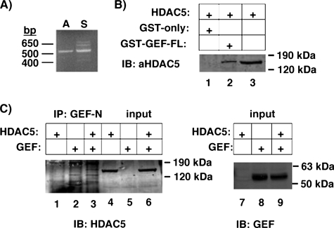
GEF can interact with HDAC5. A, cDNA from a portion of HDAC5 was expanded using RT-PCR from mouse adipose (A) or skeletal muscle (S) RNA and specific primers corresponding to an approximate 500-bp region of HDAC5. B, whole cell extracts from COS-7 cells transiently expressing HDAC were incubated with bacterially expressed GST alone or GST-GEF-FL bound to glutathione-Sepharose. Protein complexes were washed, eluted by denaturation, and analyzed by SDS-PAGE and immunoblotting (IB) for HDAC5. C, proteins from COS-7 whole cell extracts from cells overexpressing GEF alone, HDAC5 alone, or GEF and HDAC5 together were immunoprecipitated (IP) using affinity purified anti-GEF polyclonal antibodies and Protein A/G PLUS-agarose. Immunoprecipitates were eluted by denaturation and analyzed by SDS-PAGE and immunoblot for HDAC5 (lanes 1–3). Input (3%) was loaded as a comparison for HDAC levels (lanes 4–6) or GEF (lanes 7–9). FL, full-length.

