
Fig. 3.
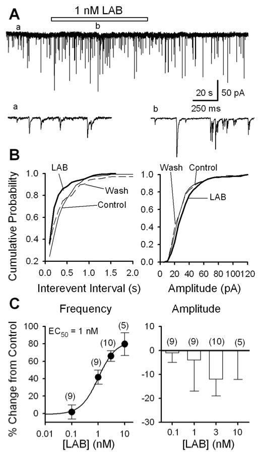
Labetalol raises the frequency of sIPSCs of PAG cells: data from mechanically dissociated neurons. A, GABAergic sIPSCs recorded before (a), during (b) and after the application of 1 nM labetalol (LAB) in a PAG cell; accelerated trace in (a) and (b) are shown. B, For the same data as A, cumulative probability plots of sIPSC interevent intervals (left: K - S test, p = 0.008, labetalol vs. control) and amplitudes (right: K - S test, p = 0.89, labetalol vs. control). C, Concentration-dependence of labetalol induced changes of the frequency (left panel, n = 5 - 10; with an EC50 of 1 nM and maximal effect of 83%) and amplitude (right panel, n = 5 - 10, p > 0.2) of sIPSCs. For all figures, the numbers in brackets are the number of neurons examined.