
Fig. 5.
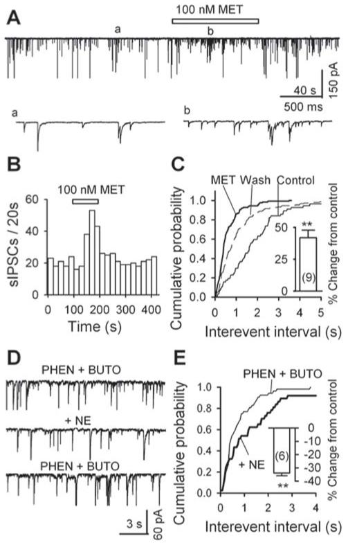
Metoprolol (MET) raises the frequency of sIPSCs: data from mechanically dissociated neurons. A, GABAergic sIPSCs recorded in a PAG cell before (a), during (b) and after the application of 100 nM MET; accelerated trace in (a) and (b). B, For the same data as in A, the time course of the enhancement of sIPSC frequency by 100 nM MET. C, Cumulative probability plots of interevent interval (left: K - S test, p < 0.001, metoprolol vs. control) and amplitude (right inset, K - S test, p = 0.037, metoprolol vs. control) of sIPSCs. Metoprolol (100 nM) significantly increased the frequency of sIPSCs (left inset: n = 9, p = 0.003). D, Norepinephrine (NE, 10 μM) decreased sIPSC frequency in the presence of phentolamine (PHEN, 10 μM), an α-adrenoceptor antagonist, and butoxamine (BUTO, 2 μM), a β2- adrenoceptor antagonist. E, cumulative probability plots of interevent interval of sIPSCs before (PHEN + BUTO) and during (+ NE) the application of 10 μM NE. Inset, the summary from 6 neurons, of the effect of norepinephrine on sIPSC frequency in the presence of phentolamine PHEN and butoxamine. ** p < 0.01, PHEN + BUTO vs. PHEN + BUTO + NE.