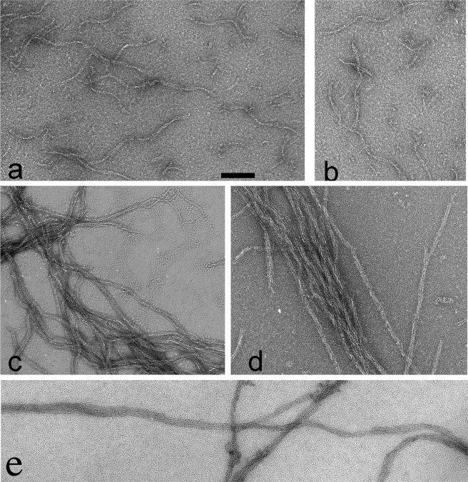
FIGURE 2.
TubZ-Ba filament structure observed by negative stain EM. a and b show TubZ-Ba filaments assembled in 100 μm GTP. The filaments mostly appear as wavy two-stranded helices, but occasional polymers have three strands (bottom 2b). c, shows TubZ-Ba filaments assembled in 20 μm GTPγS. Filaments are longer and tend to associate into bundles. In the upper right, a two-stranded filament is seen separating into single strands, d shows TubZ-Ba filaments assembled in 100 μm GTP plus 1 μm GTPγS. e shows TubZ-Ba assembled in GTP in the presence of 10% polyvinyl alcohol, a crowding agent. The TubZ-Ba concentration was 5 μm in all measurements. The scale bar is 100 nm.

