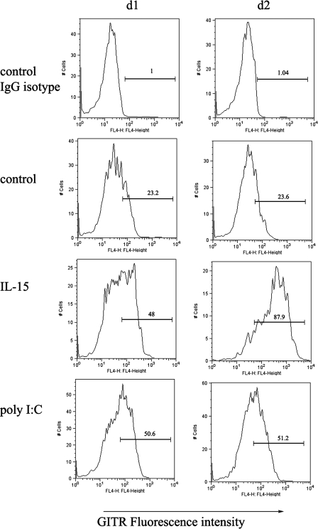
FIGURE 1.
GITR expression was up-regulated in activated NK cells. Purified PBMCs were cultured in the presence or absence of either 10 ng/ml IL-15 or 25 μg/ml poly(I-C) for 1 or 2 days. Cells were collected; stained with anti-human FITC-CD3, PE-CD56, and anti-GITR antibody; and analyzed by flow cytometry. The x axis depicts relative fluorescence intensity (expression level of GITR), whereas the y axis represents relative cell numbers. IgG isotype control indicates that the cells without treatment were stained with nonspecific isotypematched biotinylated control antibody. Control indicates that the cells without treatment were stained with 5 μg/ml biotinylated anti-human GITR antibody to show constitutive expression of GITR.

