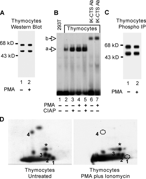
FIGURE 5.
Dephosphorylation of Ikaros in thymocytes after PMA plus ionomycin treatment. Nuclear extracts of unstimulated and PMA plus ionomycin-treated thymocytes were obtained and normalized for Ikaros concentrations by Western blot (A). B, DNA binding activities of Ikaros toward the TdT D′ regulatory elements were compared by gel shift in the absence lanes (2 and 4) or presence (lanes 3 and 5) of calf-intestine alkaline phosphatase (CIAP, 10 units). Ab, antibody. Unstimulated and PMA plus ionomycin-treated murine thymocytes were grown in the presence of [32P]orthophosphate. Ikaros proteins were immunoprecipitated (IP) from nuclear extract using anti-Ikaros antibodies (C) and used to generate two-dimensional phosphopeptide maps (D). Phosphopeptides representing novel phosphorylation sites are numbered 1–4. A star indicates a phosphopeptide representing a previously identified phosphorylation site (amino acid 63). The two phosphopeptides that were not detected in PMA plus ionomycin-treated thymocytes are indicated with a dashed circle.

