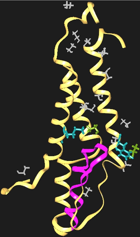
FIGURE 7.
A molecular model of the NR2A subunit M domains showing putative binding sites for ethanol at positions 637 and 823. A model of the M domains of the NR2A subunit was derived by homology modeling and is depicted as a ribbon structure. Following solvation with ethanol, MD simulations were run in which the ethanol was allowed to diffuse away to identify the most probable binding sites. The M2 segment is highlighted in purple, and Met-825 and Phe-637 are shown in light blue. Molecules of the ethanol solvent are white. Note that two ethanol molecules (light green) remain tightly associated with Phe-637 and Met-823.

