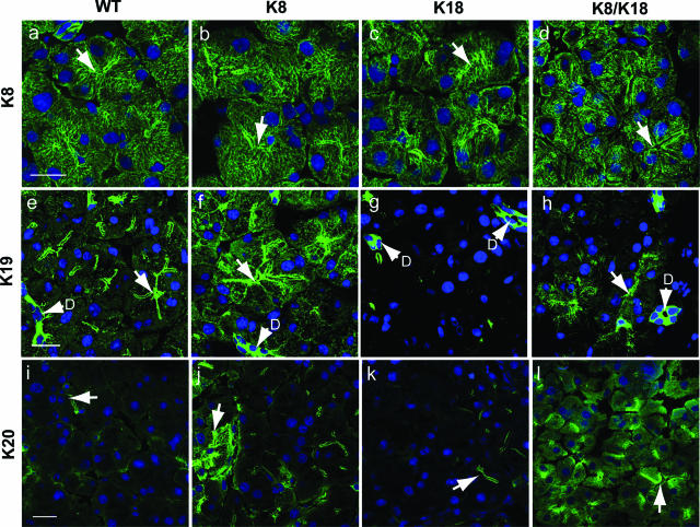
Figure 2.
Keratin immunofluorescence staining of control and keratin-overexpressor pancreata. Keratins from WT (a, e, i), K8 (b, f, j), K18 (c, g, k), and K8/K18 (d, h, l) mouse pancreata were visualized by double-immunofluorescence staining using antibodies to K8 (a–d), K19 (e–h), and K20 (i–l) and counterstained for nuclei (blue). Arrows point to examples of lumens; D, ducts. Scale bars = 20 μm.

