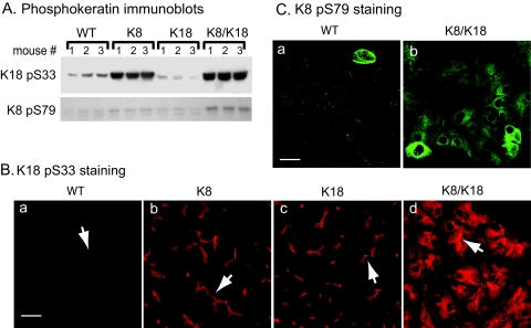
Figure 3.
Keratin phosphorylation is increased in K8 and K8/K18 mice. The level of keratin phosphorylation in WT, K8, K18, and K8/K18 mice was investigated by immunoblotting (A) as described in Figure 1 and by immunofluorescence staining using Abs to K18 pS33 and K8 pS79 (B and C, respectively). Arrows point to examples of lumens. In A, three separate mice (nos. 1 to 3) were used per genotype. Scale bars = 20 μm.

