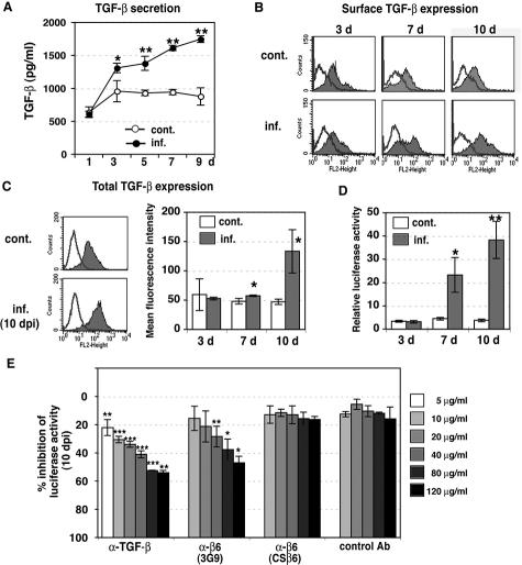
Figure 2.
Integrin αvβ6-dependent TGF-β1 activation in CMV-infected HUVECs. A: TGF-β1 production by infected HUVECs. Conditioned medium was collected from control (open circles) and infected (filled circles) HUVECs at 1 to 9 days, and TGF-β1 was quantified by enzyme-linked immunosorbent assay. Results are the mean (±SE) of three experiments done in duplicate. Asterisks indicate the amount of TGF-β1 in infected HUVECs as compared with uninfected controls (*P < 0.05, **P < 0.01). B: Surface expression of TGF-β1 in HUVECs was analyzed by flow cytometry at 3, 7, and 10 days after infection and controls (cont). Typical histograms are shown. Shaded areas represent expression of specific proteins. Lines represent isotype control. Experiments were repeated at least three times. C: Total TGF-β1 was analyzed by flow cytometry using permeabilized cells at 3, 7, and 10 days after infection (inf.) and controls (cont). Left: Typical histograms at 10 days are shown. Shaded areas represent expression of specific proteins. Lines represent isotype control. Right: Results are the mean fluorescence intensity (±SE) of three experiments. Asterisks indicate expression in infected HUVECs as compared with uninfected controls (*P < 0.05). D: TGF-β bioassay of active TGF-β produced by infected HUVECs. Equal numbers of TMLC TGF-β reporter cells, and control (cont.) or infected HUVECs (inf.) were cultured for 16 to 24 hours at 3, 7, and 10 days after infection. Relative luciferase activity in cell lysates was defined as the measured activity divided by TMLC baseline activity. Results are the mean (±SE) from 6 to 11 experiments done in duplicate. Asterisks indicate the TGF-β1 activity in infected HUVECs as compared with uninfected controls (*P < 0.05, **P < 0.001). E: Inhibition of luciferase activity in TGF-β bioassay by anti-integrin αvβ6. HUVECs infected for 10 days were co-cultured with TMLCs with anti-TGF-β neutralizing antibody (1D11); function-blocking anti-αvβ6 antibody (3G9); isotype-matched, non-function-blocking anti-αvβ6 antibody (CSβ6); or mouse IgG1 isotype control antibody (control Ab). Results are the mean (±SE) from three to five experiments done in duplicate. Asterisks indicate inhibition of TGF-β1 activation relative to untreated infected HUVECs (*P < 0.05, **P < 0.01, ***P < 0.001).

