Figure 5.
Localization of the Fluorescence Signal in PIP5K3-YFP and PIP5K3ΔM-YFP Root Hair Cells.
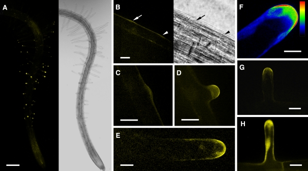
The YFP fluorescence in root hairs expressing PIP5K3-YFP and PIP5K3ΔM-YFP under the control of the PIP5K3 promoter was observed using confocal laser scanning microscopy.
(A) A three-dimensional reconstructed fluorescence image (left) and a light image (right) of a PIP5K3-YFP root at 5 d after germination growing on agar medium.
(B) Fluorescence image (left) and light image (right) of the PIP5K3-YFP root surface with an emerging bulge (arrow) and the position where a bulge was expected to emerge (arrowhead). Time-lapse fluorescence images of the same sample are shown in Supplemental Figure 5 online.
(C) An emerging bulge on the PIP5K3-YFP root hair cell surface.
(D) to (G) Elongating PIP5K3-YFP root hairs. The fluorescence intensity is shown by index colors, with red and blue for high and low intensities, respectively, in (F).
(H) An elongating PIP5K3ΔM-YFP root hair.
Bars = 0.2 mm in (A), 20 μm in (B) to (D), (G), and (H), and 10 μm in (E) and (F).

