Figure 3.
The Antiapoptotic Effect of PXR in HCT116 Cells Was Not Bile Acid Specific and May Not Rely on the Activation of Bile Acid-Detoxifying Enzymes
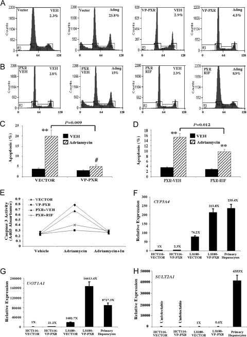
A and B, Flow cytometry analysis on adriamycin (Admg)-induced apoptosis in HCT116-VP-PXR cells (A) and HCT116-PXR cells in the presence or absence of RIF (B). Cells were treated with adriamycin (0.5 mg/ml) for 24 h before flow cytometry analysis. The percentages of apoptotic sub-G1 cell populations are labeled. Results shown are representative flow cytometry profiles from the same clones used in Fig. 2, A and B. C and D, Quantification of apoptotic cells in panels A and B, respectively. Results shown represent averages and sd derived from the same pooled clones used in Fig. 2, C and D. **, P < 0.01; #, P > 0.05. P values are compared with the Vehicle (VEH) control, or the comparisons are labeled. E, Adriamycin-induced apoptosis was measured by caspase 3 activation. Adriamycin treatment was the same as in panels A and B. In the “Adriamycin+In” lane, cells were cotreated with the caspase inhibitor Z-VAD-FMK. Results shown represent averages derived from two pooled clones. F–H, Real-time PCR analysis on the expression of bile acid-metabolizing enzymes CYP3A4 (F), UGT1A1 (G), and SULT2A1 (H) in Vector- or VP-PXR-transfected HCT116 and LS180 cells. RNA prepared from untreated primary human hepatocytes from three patients (all Caucasians, including a 45-yr-old male, a 67-yr-old female, and an 84-yr-old male) was included as positive controls.

