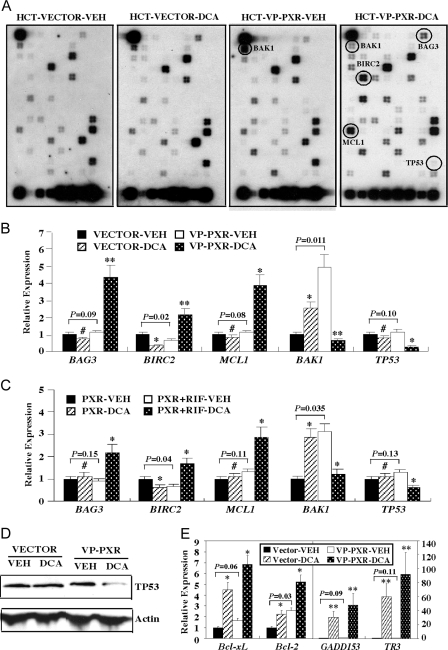
Figure 5.
Molecular Mechanism by Which PXR Inhibited DCA-Induced Apoptosis in HCT116 Cells
A, SuperArray analysis on RNA samples derived from Vector and VP-PXR cells mock treated or treated with 250 μm DCA for 2 h. Genes the expression of which notably altered are circled and labeled. B, Real-time PCR analysis of BAG3, BIRC2, MCL-1, BAK1, and TP53 mRNA expression on RNA samples described in panel A. C, Real-time PCR analysis on RNA samples derived from PXR cells mock treated or treated with 250 μm DCA for 2 h in the presence or absence of RIF treatment. The RIF (10 mm) treatment started 24 h before DCA exposure and continued until the completion of the experiment. D, Decreased expression of TP53 protein in DCA-treated VP-PXR cells as revealed by Western blot analysis. E, Real-time PCR analysis of Bcl-xL, Bcl-2 (left panel), GADD153 and TR3 (right panel) mRNA expression on RNA samples described in panel A. Results in panels B, C, and E represent averages and sd from triplicate assays. *, P < 0.05; **, P < 0.01; #, P > 0.05. P values are compared with the Vehicle (VEH) control of the same cells, or the comparisons are labeled.

