Figure 6.
Activation of PXR Inhibited Bile Acid-Induced Apoptosis and Sensitized Mice to DMH-Induced Colonic Carcinogenesis in Vivo
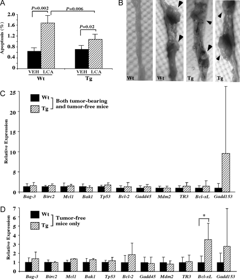
A, Wild-type (Wt, n = 3) and FABP-VP-PXR transgenic (Tg, n = 4) mice treated with vehicle or LCA enema for 5 wk. Colon tissues were then harvested and subjected to TUNEL assay for the detection of apoptotic cells. Results shown are percentages of apoptotic cells and represent averages and sd from three mice in each group. P values and comparisons are labeled. B, Representative tumor-free and tumor-bearing colon tissues derived from DMH-treated Wt and Tg mice. Mice received DMH treatment for 18 wk and were killed 12 wk after the last dose. Arrowheads indicate tumor nodules. C and D, Expression of 11 apoptosis-related genes in DMH-treated Wt and Tg mice when both tumor-bearing and tumor-free mice (C) or only the tumor-free mice (D) were included in the analysis. *, P < 0.05, compared with the Wt mice. VEH, Vehicle.

