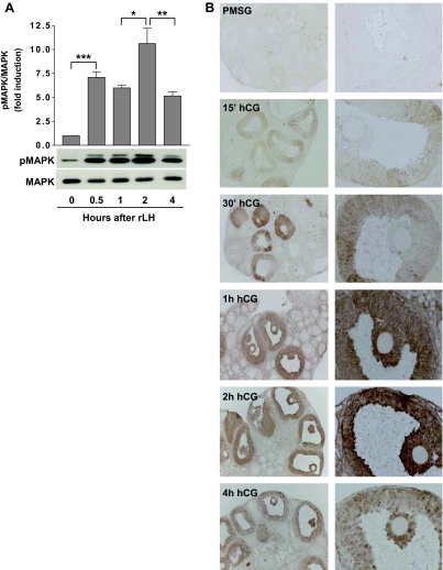
Figure 3.
Time Course of MAPK Phosphorylation in Vitro and in Vivo
A, Time course of MAPK activation in POFs. MAPK phosphorylation was examined by Western blot analyses using protein extracts from POFs cultured in the presence of rLH (5 IU) for 0, 0.5, 1, 2, and 4 h. The ratio of phosphorylated MAPK to total MAPK was determined for each time point, and the relative values are expressed as fold induction over control. Representative blots are provided. Data are shown as the mean ± sem of five separate experiments. *, P < 0.05; **, P < 0.01; ***, P < 0.001. B, Immunolocalization of activated MAPK in mouse ovaries. Phosphorylated MAPK was localized in hormone-primed ovaries by immunohistochemical analyses using an anti-phospho-p44/42 MAPK antibody. No signal above background was observed in PMSG-primed mouse ovaries. After 15–30 min hCG stimulation, staining for phosphorylated MAPK was detected predominantly in mural granulosa cells of POFs. At 1 h hCG, signal was evident in both mural granulosa and cumulus cell compartments and remained elevated after 2 h. Theca cells of preantral and antral follicles were also positive at these times. After 4 h, signal was still detected in cumulus cells and appeared decreased in granulosa cells of POFs.

